


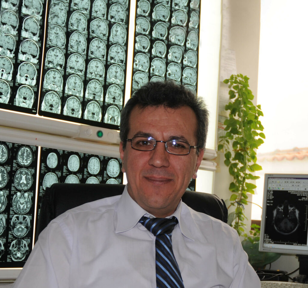
by Dr. Panagiotis Toulas
The brain is the center of our nervous system. It is a complex organ that controls the body’s function and coordinates its organs to function in symphony.
The importance and the delicacy with which the brain functions require a special protection, that is, inside the cranial cavity. In other words, this cavity is a hard bony box called, the cranium, the inside of which is lined with hard membranes called the meninges. Furthermore, within the cranial cavity, there is the cerebrospinal fluid (CSF) that flows in and around the brain.
The human brain is extended and controls a supportive “skeleton” which consists of the neuroglia or glia and about 100 billion neurons. The neurons or nerve cells are connected and communicate with each other by creating a functional type of electrical circuit with numerous neural extensions and interactions.
Cell communication is achieved by the synapses which are functional neural connections with chemical mediators (e.g., neurotransmitters and electrolytes). In other words, the synapse is a small gap between two neurons, where nerve impulses are relayed by a neurotransmitter from the sending neuron to the one of the receiving neuron which is how the cell communication is achieved.
Macroscopically, when one examines the brain, they may notice that it consists of symmetrical parts (which are connected to each other) and distinct anatomical areas such as the two cerebral hemispheres; The cerebellum and the brain stem. The organization of each part of the brain to perform simple or more complex functions led us to describe the various areas responsible for these functions, which may or may not work in cooperation with each other.
Many studies have confirmed that the cerebral hemispheres have specific parts for specific functions. In other words, the occipital lobes are responsible for visual perception, the parietal lobes are responsible for processing somatosensory information from the body and they also are the centers of logical thinking and speech. The frontal lobes are important for voluntary movement, expressive language and for managing higher level executive functions, while the temporal lobes are primarily responsible for interpreting sounds, and recognizing and using languages, and they also contribute to object recognition and creation of new and long term memories.
There are also specific areas which differ either functionally or anatomically. Nevertheless, they are part of many lobes and thus participate in many functions, e.g., basal ganglia, limbic system, etc.
The brain being an important regulatory organ responsible for the survival of the organism, is supplied with blood by many large blood vessels which interact with each other. Therefore, should one such vessel undergoes damage, the rest of them will continue the blood supply. After all, the brain, in proportion to its volume, receives a significant amount of blood.
Imaging Examination
On the occasion of World Brain Day, celebrated every year on July 22, which was an initiative of the World Federation of Neurology, we will attempt to summarize the technological progress recorded in recent decades in terms of imaging tests, which has contributed the most to further understanding of brain function.
Computed Tomography (CT), is the anatomical depiction tool of the brain. Magnetic Tomography, which has dynamically entered daily practice in the last 20 years or so, offers us more detailed images of the brain, without the use of ionizing radiation. Additionally, we are able to monitor the condition of the brain’s vessels without the patient having to undergo invasive angiographic examinations and the use of catheters.
Magnetic Resonance Imaging (MRI) and Magnetic Spectroscopy (MRS) offered us the way of measuring the substances (metabolites) of the healthy brain and potential brain damage.
Therefore, a biopsy can be avoided, which presupposes the performance of – even a small – operation. In recent years, the so-called Functional Magnetic Tomography (Functional MRI-fMRI) has also made its debut into the clinical daily practice of Magnetic Resonance Imaging. Functional MRI-fMRI identifies the exact area of the brain that performs a specific function (e.g., which area moves the hand, foot, tongue, or which area is responsible for speech, vision, etc.). The study of these elementary functions led over time to the study of more complex functions (e.g., thinking, feelings, etc.).
The Polydynamo Center of the BIOIATRIKI Healthcare Group in Cyprus is equipped with a 3 Tesla MRI, the first of its kind with artificial intelligence in Cyprus and Greece, and only the third that Siemens has installed in Europe. That device has also interconnection capabilities with international databases and thus offers the most valid and accurate diagnostic results. This is a state-of-the-art system with the most spacious examination ring and innovative special applications that enable specialized neuro-imaging examinations such as MR spectroscopy, MR tractography, MR perfusion and MR diffusion, and many more.
*Neuroradiologist, BIOIATRIKI Healthcare Group Imaging Director, Scientific Associate of the Radiology Research Unit of the National and Kapodistrian University of Athens.